Back Of Neck Anatomy Muscles - A collection of anatomy notes covering the key anatomy concepts that medical students need to learn.. Muscles and ligaments work together to support the spine, hold it upright, and control movement during rest and activity. 21 muscles of the neck: There are four pairs of muscles that are responsible for chewing movements or mastication. Is the only cutaneous muscle in human body (under the skin) attachments: Some important structures contained in or passing through the neck include the seven cervical vertebrae and enclosed spinal cord, the jugular veins and carotid arteries, part of the esophagus, the larynx.
Muscles of the neck are described separately from the compartments. The muscles of the neck run from the base of the skull to the upper back and work together to bend the head and assist in breathing. By the middle line of the back is a longitudinal groove back (sulcus dorsi). Intermediate back muscles and c. A collection of anatomy notes covering the key anatomy concepts that medical students need to learn.
Topographic Anatomy Of The Back The Lecturio Medical Online Library from d3uigcfkiiww0g.cloudfront.net We will attempt to provide a simplified overview of this complex anatomy. From the mastoid process of the temporal bone goes the muscle that inserts into the inner. The splenius muscles originate at the midline and run laterally and. There are a bunch of muscles in the back of your neck which help lift your head up. The muscles of the back that work together to support the spine, help keep the body upright and allow twist and bend in many directions. Some important structures contained in or passing through the neck include the seven cervical vertebrae and enclosed spinal cord, the jugular veins and carotid arteries, part of the esophagus, the larynx. Working in pairs on the left and. Along it are easily palpable spinous processes by palpation of the cervical vii and all lying.
Anterior muscles of the neck.
21 muscles of the neck: The muscles of the head and neck help us perform many important actions such as movement the muscles of the pelvis, hip and buttock anatomical chart shows how each muscle in this area of the extensor muscles are connected to the back of the spine and allow a person to stand and to lift. Bones of the neck picture. Inserts on to the humerus. The extrinsic muscles that are associated with upper extremity and shoulder movement, and the they laterally flex, rotate, and extend your head and neck. Neck muscles are bodies of tissue that produce motion in the neck when stimulated. From the mastoid process of the temporal bone goes the muscle that inserts into the inner. The posterior muscles of the neck are primarily concerned with head movements, like extension. Brings down corners of the mouth, expressing.
Rectus capitis, longus capitis, longus colli. Is the only cutaneous muscle in human body (under the skin) attachments: Along it are easily palpable spinous processes by palpation of the cervical vii and all lying. Back muscles are arranged in several layers, so they are divided into deep and superficial, which, in turn, are arranged in two layers. Muscles are named according to their shape, location, or a combination.
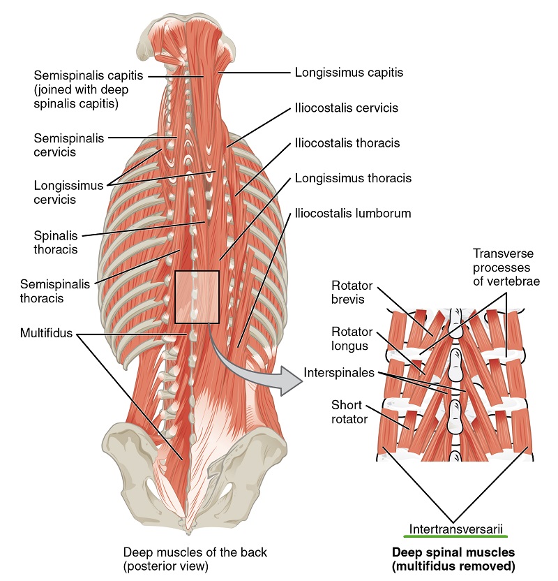
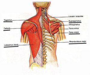
Muscle Knots In Your Back And Neck Elements Chiropractic Clinic from www.elementschiropractic.com Bones of the neck picture. There are several individual muscles within the back anatomy, and it's important to take a quick look the image below to shows all the major back muscles (as well as some neck muscles) There are a bunch of muscles in the back of your neck which help lift your head up. Back muscles are arranged in several layers, so they are divided into deep and superficial, which, in turn, are arranged in two layers. Here the extrinsic back muscles are classified into logical subgroups to facilitate knowledge. Neck, in land vertebrates, the portion of the body joining the head to the shoulders and chest. Tutorials and quizzes on the anatomy and actions of the back muscles (iliocostalis, longissimus, spinalis, multifidus, and quadratus lumborum), using interactive animations, diagrams, and illustrations. 21 muscles of the neck:
Brings down corners of the mouth, expressing.
From the mastoid process of the temporal bone goes the muscle that inserts into the inner. The major muscles of the back, from superficial to deep are divided in three groups: The back muscles stabilize and move the vertebral column, and are grouped according to the lengths and direction of the fascicles. Neck muscles are bodies of tissue that produce motion in the neck when stimulated. Extrinsic, intermediate and intrinsic muscles. Neck muscles help support the cervical spine and contribute to movements of the head, neck, upper back, and posterior longitudinal ligament (pll). Brings down corners of the mouth, expressing. Sternohyoid, sternothyroid, thyrohyoid, omohyoid anterior vertebral muscles: The anterior and middle scalenes originate from the transverse processes of certain cervical vertebrae and attach to the first rib.
There are four pairs of muscles that are responsible for chewing movements or mastication. Back muscles are divided into two specific groups: Muscles of the head & neck | anatomy model. The muscles of the head and neck help us perform many important actions such as movement the muscles of the pelvis, hip and buttock anatomical chart shows how each muscle in this area of the extensor muscles are connected to the back of the spine and allow a person to stand and to lift. The back muscles stabilize and move the vertebral column, and are grouped according to the lengths and direction of the fascicles.
Your Back Neck Muscles What They Look Like Bizlinks from bizlinks.files.wordpress.com This article describes the anatomy of the head and neck of the human body, including the brain, bones, muscles, blood vessels, nerves, glands, nose, mouth, teeth, tongue, and throat. Included are views of the back of the neck, short muscles of the neck, prevertebral muscles. There are a bunch of muscles in the back of your neck which help lift your head up. The back muscles can be three types. There are four pairs of muscles that are responsible for chewing movements or mastication. Is the only cutaneous muscle in human body (under the skin) attachments: Figure 11.13 muscles of the anterior neck the anterior muscles of the neck facilitate swallowing and speech. Muscles of the head & neck | anatomy model.
The anterior and middle scalenes originate from the transverse processes of certain cervical vertebrae and attach to the first rib.
There are several individual muscles within the back anatomy, and it's important to take a quick look the image below to shows all the major back muscles (as well as some neck muscles) Bodies have two kinds of splenius muscles: Figure 11.13 muscles of the anterior neck the anterior muscles of the neck facilitate swallowing and speech. Rectus capitis, longus capitis, longus colli. The muscles of the back that work together to support the spine, help keep the body upright and allow twist and bend in many directions. Back muscles are divided into two specific groups: Digastric, mylohyoid, geniohyoid, stylohyoid infrahyoid muscles: Cervical spine anatomy is quite complex. Included are views of the back of the neck, short muscles of the neck, prevertebral muscles.
Berbagi :
Posting Komentar
untuk "Back Of Neck Anatomy Muscles - A collection of anatomy notes covering the key anatomy concepts that medical students need to learn."


Posting Komentar untuk "Back Of Neck Anatomy Muscles - A collection of anatomy notes covering the key anatomy concepts that medical students need to learn."